The venous system in our legs consists of a complex network of vessels designed to return blood to the heart against gravity. With over 40 million Americans affected by venous disorders and women experiencing these conditions twice as often as men, understanding the different types of veins in the legs is essential for maintaining good vascular health.
Main Types of Veins in Legs
The leg’s venous system is organized into three distinct categories, each with specific functions and characteristics:
1. Superficial Veins
Superficial veins run close to the skin’s surface and are often visible beneath the skin. These include:
- Great Saphenous Vein (GSV) – The longest vein in the human body, extending from the foot to the groin along the inner leg. Contains 10-20 valves that prevent blood from flowing backward.
- Small Saphenous Vein (SSV) – Runs along the back of the leg from the ankle to the knee, connecting to the popliteal vein.
- Accessory Saphenous Veins – Smaller tributary veins that connect to the main saphenous veins, including anterior and posterior accessory saphenous veins.
These superficial veins are more prone to disorders like varicose veins and spider veins due to their location and less structural support.
2. Deep Veins
Deep veins are located beneath muscles and handle approximately 90% of blood flow from the legs. Key deep veins include:
- Femoral Vein – The main deep vein in the thigh that collects blood from smaller veins and transports it upward toward the heart.
- Popliteal Vein – Runs behind the knee in the popliteal fossa, receiving blood from the lower leg and connecting the tibial veins to the femoral vein.
- Tibial Veins – Include anterior, posterior, and peroneal veins that run along the shin bones, collecting blood from the foot and calf.
Deep veins work with the calf muscle pump to push blood against gravity. Issues with these veins can lead to serious conditions like deep vein thrombosis (DVT).
3. Perforator Veins
Perforator veins create crucial connections between the superficial and deep venous systems:
- Medial Calf Perforators (Cockett’s perforators) – Located along the medial lower leg
- Posterior Tibial Perforators – Found behind the tibia
- Boyd’s Perforators – Positioned below the knee
- Hunterian Perforators – Located in the mid-thigh region
These veins pierce through muscle fascia, allowing blood to flow from superficial veins into the deep veins. When perforator valves fail, blood flows backward, potentially causing varicose veins and other venous disorders.
How Blood Flows in Leg Veins
Blood flow in the types of veins in the legs relies on three key mechanisms:
- Muscle Pump Action – Contractions of calf and thigh muscles compress veins, pushing blood upward
- Respiratory Pump – Changes in abdominal pressure during breathing help draw blood toward the heart
- Venous Valves – One-way valves prevent backward flow, maintaining forward movement of blood
When these mechanisms function properly, blood efficiently returns to the heart. However, when valve function declines, venous insufficiency can occur, causing blood to pool in leg veins.
Common Disorders Affecting Different Types of Veins in the Legs
Superficial Vein Disorders
- Varicose Veins – Enlarged, twisted veins that appear blue or purple and often bulge beneath the skin
- Spider Veins – Smaller, dilated blood vessels appearing as thin, web-like red, blue, or purple lines
- Reticular Veins – Medium-sized blue veins visible beneath the skin but not raised
Deep Vein Disorders
- Deep Vein Thrombosis (DVT) – Blood clots form in deep veins, causing pain, swelling, warmth, and redness
- Post-thrombotic Syndrome – Long-term complications following DVT, including chronic pain and swelling
Perforator Vein Disorders
- Incompetent Perforator Veins – When perforator valves fail, causing increased pressure in superficial veins
- Venous Ulcers – Often develop near incompetent perforators, particularly around the ankle
Diagnosing Issues in Different Types of Veins in the Legs
Diagnostic ultrasound is the gold standard for assessing the types of veins in the legs. This non-invasive imaging technique:
- Creates detailed images of vein structure
- Evaluates blood flow patterns and valve function
- Identifies areas of reflux or blockage
- Distinguishes between superficial and deep vein issues
For different vein types, specific diagnostic protocols are employed:
- Superficial Veins – Ultrasound assessment focuses on the great and small saphenous veins
- Deep Veins – Compression ultrasound tests vein compressibility to identify potential clots
- Perforator Veins – Specialized ultrasound techniques locate and assess the function of connecting vessels
Treatment Options for Various Types of Veins in the Legs
Superficial Vein Issues
- Sclerotherapy – Injections that cause small varicose and spider veins to collapse and fade
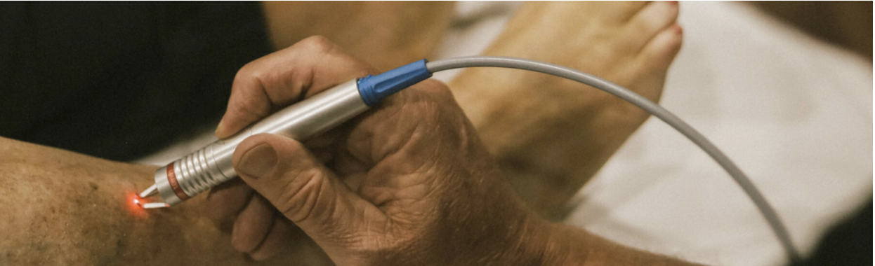
- Superficial Laser Treatment – Targets spider veins with precisely directed energy
- Microphlebectomy – Removes varicose veins through tiny incisions using specialized hooks
Deep Vein Issues
- Anticoagulation Therapy – Blood-thinning medications to prevent clot growth
- Thrombolysis – Breaking down existing clots using specialized medications
- Venous Stenting – Placing mesh tubes to keep compressed veins open
For All Types of Veins in Legs
- Compression Therapy – Compression stockings provide external support to vein walls and work with the calf muscle pump to minimize swelling and pain
- Lifestyle Modifications – Regular exercise, weight management, and avoiding prolonged sitting or standing
Preventing Problems in Different Types of Veins in the Legs
To maintain healthy veins in your legs:
- Stay active with regular walking or swimming to enhance calf muscle pump function
- Take breaks from prolonged sitting or standing to promote circulation
- Maintain a healthy weight to reduce pressure on leg veins
- Elevate legs when resting to assist venous return
- Consider wearing compression stockings, especially if at risk for venous issues
- Stay hydrated and limit high-sodium foods to reduce water retention and swelling
Conclusion
Understanding the different types of veins in the legs—superficial, deep, and perforator—is crucial for recognizing potential issues and seeking appropriate care. Each type plays a vital role in returning blood to the heart, and problems in any part of this system can lead to various venous disorders.
When experiencing symptoms like leg discomfort, swelling, or visible vein changes, early detection through diagnostic ultrasound can make all the difference in treatment outcomes. With modern treatment options ranging from compression therapy to minimally invasive procedures, maintaining healthy leg veins is achievable.
Frequently Asked Questions
What are the main types of veins in the legs? The main types include superficial veins (Great and Small Saphenous), deep veins (femoral, popliteal, tibial), and perforator veins that connect the two systems.
Which types of veins in the legs are most prone to varicose veins? Superficial veins, particularly the Great Saphenous Vein, are most commonly affected by varicose veins due to their location closer to the skin surface and less muscular support.
How do the different types of veins in the legs work together? Blood flows from superficial veins through perforator veins into the deep veins, which carry most of the blood back to the heart. This system relies on one-way valves and muscle contractions to move blood against gravity.
Can problems in one type of vein affect other types of veins in the legs? Yes, issues like valve failure in perforator veins can cause increased pressure in superficial veins, leading to varicose veins. Similarly, deep vein thrombosis can affect overall circulation in the leg.
Which types of veins in the legs are examined during a vascular ultrasound? A comprehensive vascular ultrasound examines all three types of veins in the legs—superficial, deep, and perforator veins—to assess structure, blood flow, and valve function throughout the entire venous system.